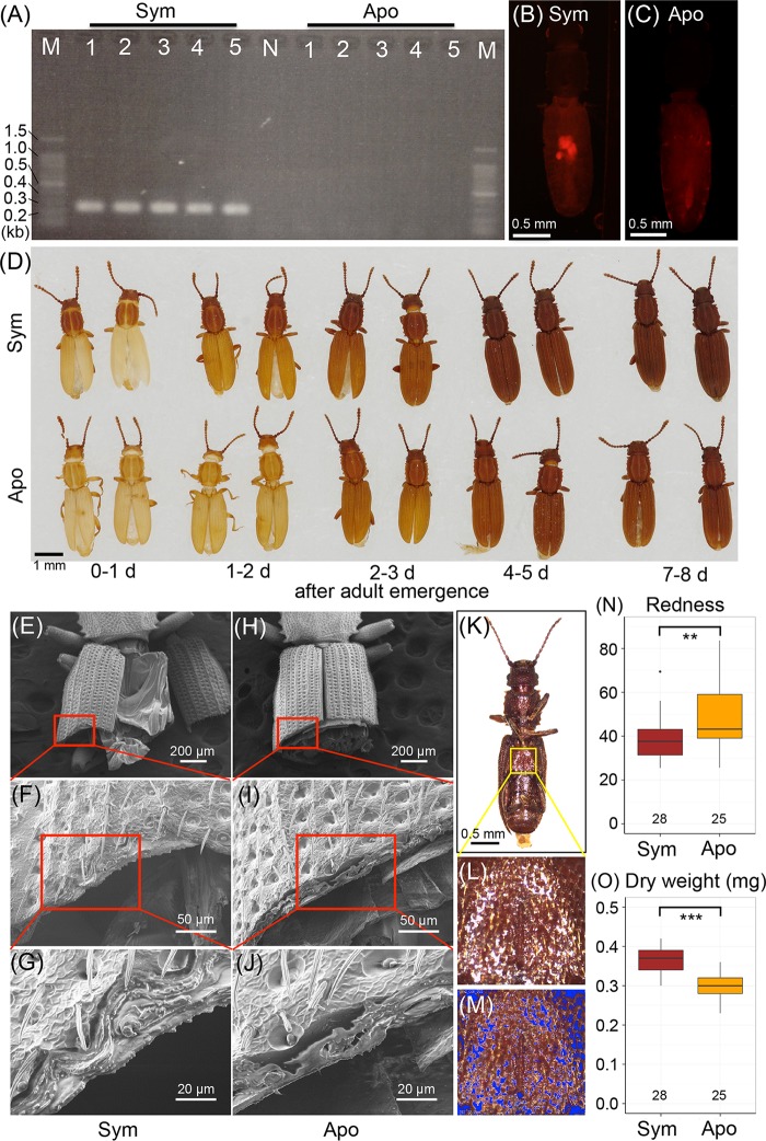
FIG 8 .
Effects of symbiont removal on adult phenotypes of O. surinamensis. (A) Diagnostic PCR detection of the symbiont from adult insects of the control symbiotic line (Sym) and the antibiotic-treated aposymbiotic line (Apo). Each lane in lanes 1 to 5 represents a DNA sample from a single insect. N and M represent the no-template control and DNA size markers, respectively. (B and C) Fluorescence in situ hybridization targeting 16S rRNA of the symbiont in adult females of the symbiotic strain (B) and the aposymbiotic strain (C). (D) Cuticle pigmentation after adult emergence of the symbiotic insects (upper row) and the aposymbiotic insects (lower row). (E to J) Scanning electron microscopic images of sectioned elytra of the symbiotic adult insects (E to G) and the aposymbiotic adult insects (H to J). Panels F and I are magnified images of panels E and H, as indicated by red rectangles; panels G and J are magnified images of panels F and I. (K to M) Process of quantifying redness of cuticle. From each of the ventral images of adult insects (K), a square area was extracted from the metasternum. On the square image (L), the pixels whose brightness was either above the top 10% or below the bottom 10% were masked in blue (M) and excluded from the analysis to minimize the effects of highlights and shadows. Then, RGB values for all (n) pixels were measured and averaged to obtain the redness index by Σ[R − mean (R, G, B)]/n. (N) Comparison of cuticle redness between the symbiotic insects and the aposymbiotic insects older than 2 weeks after adult emergence. (O) Comparison of dry body weight between the symbiotic insects and the aposymbiotic insects older than 2 weeks after adult emergence. In panels N and O, asterisks indicate statistically significant differences by t test: **, P < 0.01; ***, P < 0.001.

