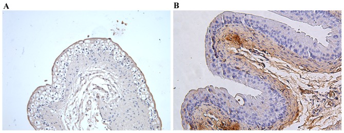
Figure 3.
Representative immunohistochemical images of ZO-1 protein. For immunohistochemical analysis, tissue sections were incubated with primary antibody anti-ZO-1 overnight at 4°C. (A) In control mice, ZO-1 was localized to superficial umbrella cell layer at the interendothelial junctions in most samples. (B) In the ketamine group, ZO-1 located in the cytoplasm and not organized into tight junction structures, or absent. Magnification, ×200; scale bar, 100 µm. ZO-1, zonula occludens-1.

