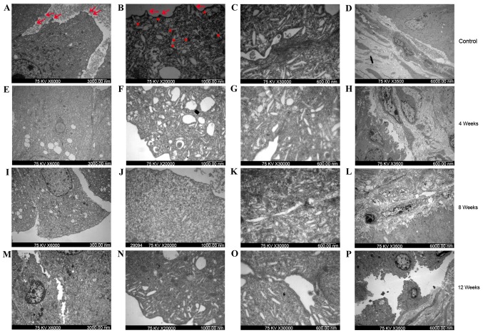
Figure 4.
Bladder uroepithelium and lamina propria from control and ketamine-treated mice detected using transmission electron microscopy. Bladder samples from (A-D) control mice and ketamine-treated mice following (E-H) 4, (I-L) 8 and (M-P) 12 weeks of treatment. (A, E, I and M) Cell-cell connections (magnification, ×6,000; scale bar, 3,000 nm). The gap between cells in the ketamine-treated groups (E, I and M) was wider than that observed in the control group (A). (B, F, J and N) Apical membrane of umbrella cells (magnification, ×20,000; scale bar, 1,000 nm). The control group (B) exhibited a normal appearance, with numerous subapical vesicles (asterisks) and raised microplicaes (arrows). Rare subapical vesicles and raised microplicaes were observed in the ketamine treatment groups (F, J and L). (C, G, K and O) Junctional complexes (magnification, ×30,000; scale bar, 600 nm). Control group (C) exhibited distinct tight junctional complexes, whereas the ketamine treatment groups (G, K and O) exhibited broken junctional complexes. Cells from the bladders of mice treated with ketamine for 4 weeks lost their cytoplasmic density. (D, H, L and P) Bladder lamina propria (magnification, ×3,500; scale bar, 6,000 nm). The control group and 4-week ketamine treatment group exhibited normal appearance of lamina propria. Vascular endothelial cells of the 8-week ketamine treatment group exhibited cell body shrinkage, cytoplasm density increase and chromatin condensation. The 12-week ketamine treatment group exhibited discontinuity in the umbrella cell layer and denuded epithelium.

