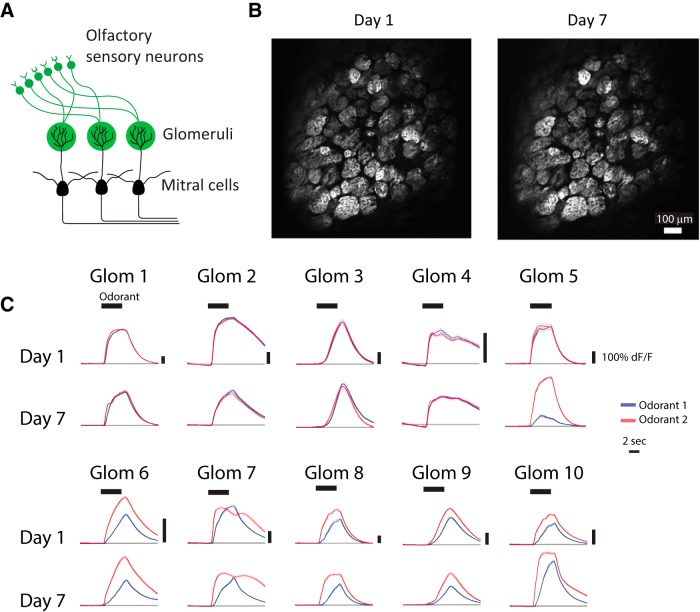
Figure 2.
Imaging glomerular odor responses during training. A, Schematic of the olfactory bulb. Two photon imaging of glomerular responses was performed in OMP-tTA::tetO-GCaMP6s mice, in which OSNs express GCaMP6s. B, An example of a typical glomerular field of view on the first day of imaging (day 1) and 6 d later (day 7). C, Examples of odorant responses (mean ± SEM) from individual glomeruli. Responses to the odorant 1 (S+, rewarded odorant) are shown in blue, and responses to odorant 2 (S-, unrewarded odorant) are shown in black. Odorant period is indicated by the thick horizontal black bar.

