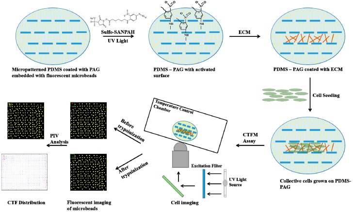
Figure 3.
The schematic illustration for the making of PDMS microchannels with a polyacrylamide gel (PAG) coating for cell traction force microscopy measurement (Top views). The micropatterned PDMS is coated with PAG embedded with a thin layer of fluorescent microbeads. A UV-activated heterobifunctional cross linker, sulfo-SANPAH, is applied for ECM coupling. Cells are then seeded onto the activated surface for further study with cell traction force microscopy. After obtaining a pair of fluorescent images of the same frame before and after trypsinization, the deformation of the elastic substrate is determined and used for the CTF computation.

