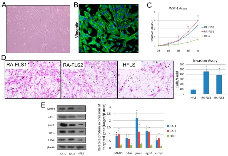
Figure 1.
Isolation and identification of fibroblast-like synoviocytes (FLS) from synovial tissues of rheumatoid arthritis (RA) patients. (A) Microscope photo of FLSs isolated from human synovial tissue at passage 3 (100×). (B) Immunofluorescence analysis of the isolated FLSs using anti-Vimentin antibody and DAPI. (C) WST-1 assay, measuring the activity of mitochondrial dehydrogenases, was performed following the manufacturer’s instructions at 0-, 1-, 2-, 3- and 4-day time points. Error bars represent standard deviation (SD) of the mean. * p < 0.05 versus the normal human FLS line (HFLS). (D) Cell invasion was determined using a Transwell assay as described in the Materials and Methods Section. Microscopic image of migrated cells is shown. Original magnification: 200×. Diagrams of migrating cells from the groups are shown, which were obtained from more than three independent experiments. * p < 0.05 versus HFLS. (E) Total cell lysates of RA–FLS1 and 2, and HFLS, were examined by Western blotting. Diagrams of the relative gray value are shown, which were obtained from more than three independent experiments. * p < 0.05 versus HFLS.

