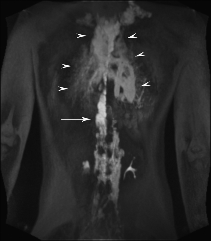
Fig. 4.

DCRML imaging of the patient with GLA, and progressive deterioration of pulmonary function and hemoptysis demonstrated dilated TD (white arrow) and abnormal pulmonary lymphatic perfusion that originates in the distal TD toward lung parenchyma (white arrowheads).
