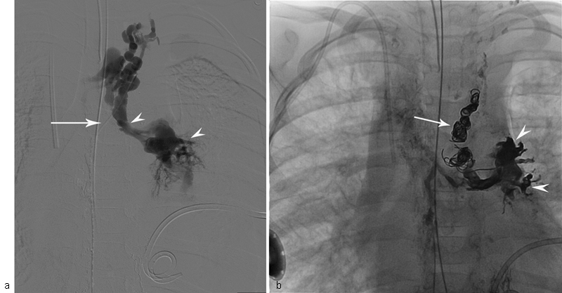
Fig. 6.

Fluoroscopic image of the injection of the contrast into TD through microcatheter (white arrow) of the patient with KL, and progressive deterioration of pulmonary function and left pleural effusion. ( a ) Image demonstrates abnormal pulmonary lymphatic flow from the TD into the lung parenchyma (white arrowheads). ( b ) Fluoroscopic image of the same patient shows endovascular coils (white arrow) and glue cast (white arrowheads) in the pulmonary lymphatics.
