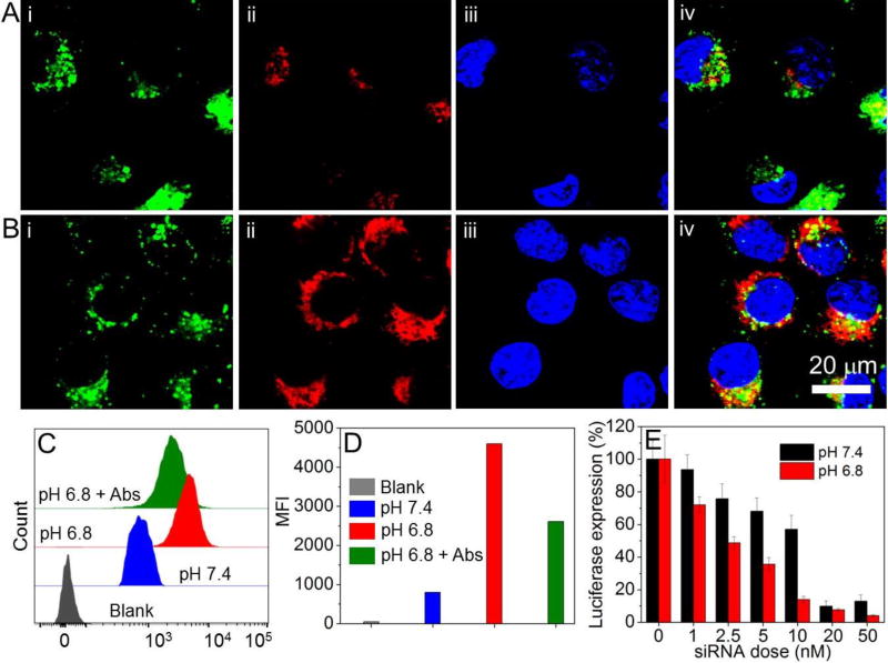
Figure 3.
(A, B) CLSM images of Luc-HeLa cells incubated with DY677-siRNA-loaded TCPA2-NPs at pH of 7.4 (A) or 6.8 (B) for 2 h. Endosomes are stained by lysotracker green, and nuclei were stained by Hoechst 33342. (i) Endosomes with green fluorescence; (ii) DY677-siRNA with red fluorescence; (iii) Nuclei with blue fluorescence; and (iv) Overlap of (i), (ii) and (iii). (C) Flow cytometry profile and (D) mean fluorescence intensity (MFI) of Luc-HeLa cells incubated with DY677-siRNA-loaded TCPA2-NPs at different pHs for 2 h, and the cells incubated with integrin αvβ3 and αvβ5 antibodies for 15 min followed by incubation with the DY677-siRNA-loaded TCPA2-NPs at pH 6.8 (pH 6.8 + Abs) for 2h. (E) Luc expression in Luc-HeLa cells treated with siLuc-loaded TCPA2-NPs at different pHs.

