
Fig. 6.
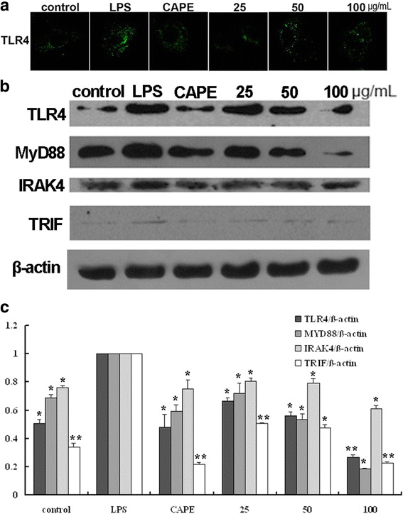
EECP and CAPE regulated the levels of TLR4, MyD88, IRAK4 and TRIF. a Cells were treated with EECP and CAPE for 24 h, respectively. Cells were stained with anti-TLR4 antibody. Immunofluorescence graphs showed a decrease of TLR4 level. b The expression of TLR4, MyD88, IRAK4 and TRIF in LPS-stimulated MDA-MB-231 cells were detected by western blotting at 48 h. c Quantification of relative expression of TLR4, MyD88, IRAK4 and TRIF in LPS-stimulated MDA-MB-231 cells. (* P < 0.05, ** P < 0.01 vs control, n = 3). Data are means ± S.E.M