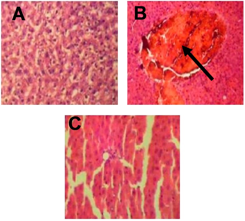
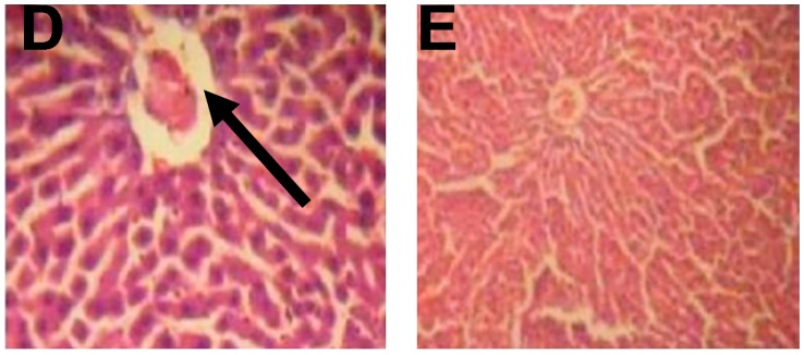
Figure 1.
Histological examination of rat livers stained with hematoxylin and eosin (H&E). (A) Control: showing normal hepatic architecture with no lesions or abnormalities; (B) CCl4: showing congestion in the central vein associated with the infiltration of inflammatory cells; (C) T1: showing mild hepatocytes necrosis and mononuclear cellular infiltration; (D) T2: showing mild portal tract and lobular chronic inflammation with focal hepatocyte destruction; (E) T3: showing that improved hepatic architecture, inflammatory cell infiltration, and hepatocytes necrosis were hardly detected (×400).


