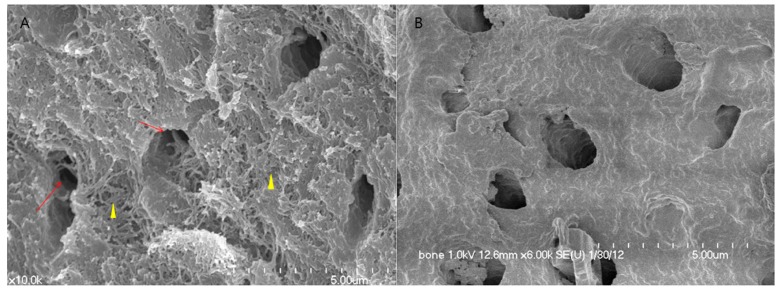
Figure 1.
SEM image of DDM. (A) Surface of DDM. Cross-sectioned tubules showing exposed outer peritubular (arrow) and intertubular collagen fibrils (arrowhead) after demineralization; (B) Surface of DDM/rhBMP-2. rhBMP-2 (0.2 mg/mL, Cowell BMP, Busan, Korea) was fixed to DDM. Structurally, the exposed collagen fibrils are impregnated with the protein solution around the dentinal tubules and collagen matrix.

