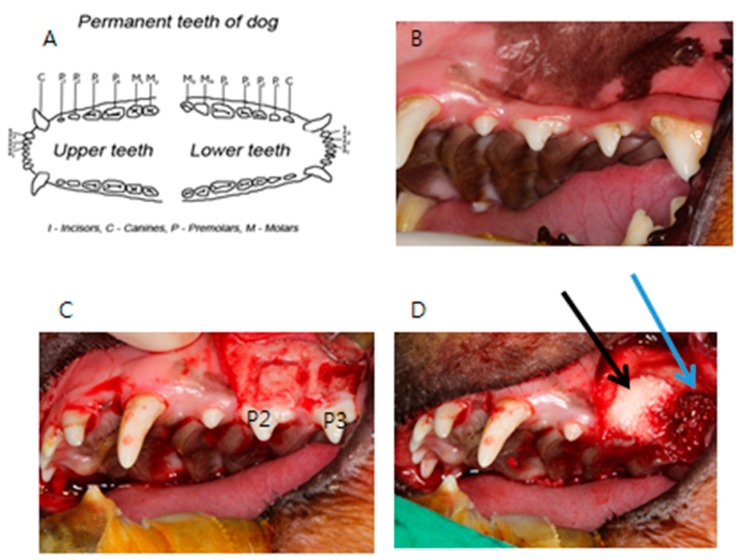
Figure 2.
Photograph of the animal experiment. (A) Permanent teeth of the beagle; (B) Intraoral photograph of the beagle; (C) Unilateral maxillary second (P2) and third premolars (P3) were extracted and mucoperiosteal flaps were elevated. The rectangular bone defects measuring 5 mm × 8 mm on the second and third premolars were made; (D) Autogenous bone was grafted onto one of two bone defects (control group, blue arrow), and human DDM fixed with rhBMP-2 was grafted onto the other (experimental group, black arrow).

