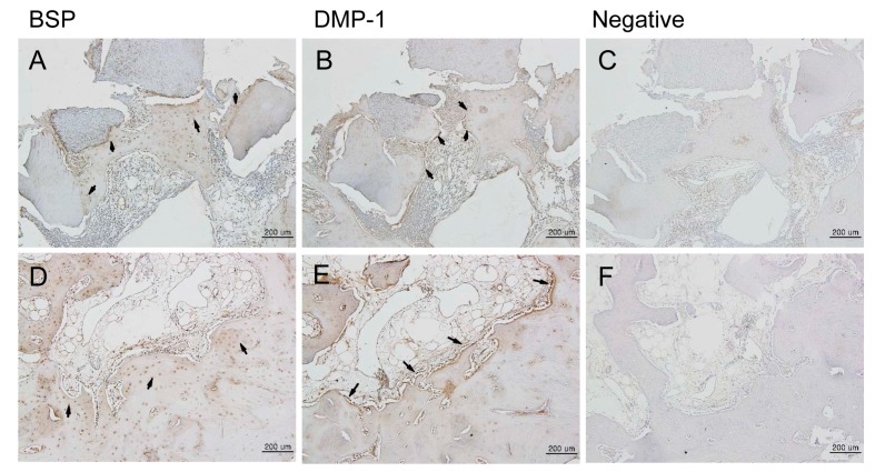
Figure 6.
Immunohistochemical analysis of new bone formation at the bone defect area after four weeks (100×). BSP, DMP-1 proteins were expressed in the area of new bone formation after four weeks. (A–C) Experimental group (DDM fixed with rhBMP-2); (D–F) Control group (autogenous bone graft). New bone tissues were immunostained with anti-BSP (A,D) and anti-DMP-1 (B,E). Negative controls (C,F) included no primary antibody. (A,D) BSP expression was detected by immunohistochemistry in the area of new bone formation. BSP was expressed by osteocytes (arrows). (B,E) DMP-1 was expressed by osteoblasts (arrows). Scale bars: 200 μm.

