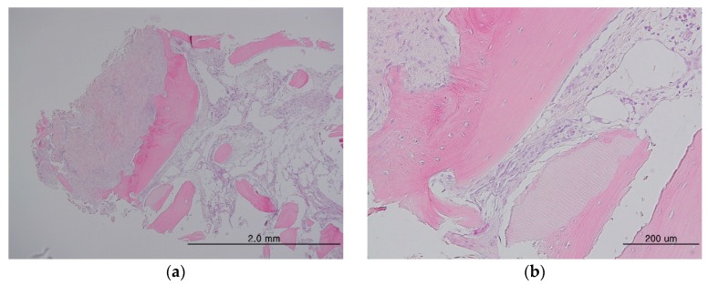
Figure 8.
Histology specimens from the maxilla and sinus (a) showing dense fibrous connective tissue on the left of the newly-formed bone, whereas tissue rich in angiogenesis can be seen on the other side of the new bone. Higher magnification showing newly-formed bone with embedded osteocytes surrounding the particles and direct contacts between the bone and particle with the vasculature (b). No inflammatory cellular infiltration can be observed.

