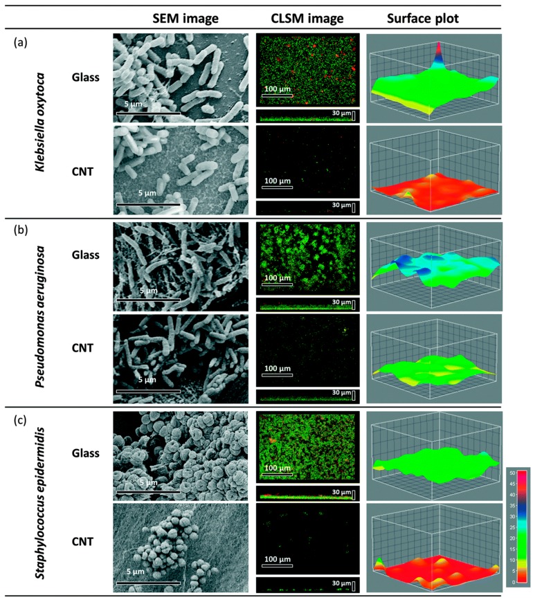
Figure 3.
Scanning Electron Microscope (SEM), Confocal Scanning Laser Microscopy (CLSM) and surface plots of biofilm formation of K. oxytoca (a); P. aeruginosa (b) and S. epidermidis (c) on multi-wall carbon nanotubes (MWCNT) (tube length 540 μm) and glass control. Reprinted with permission from Reference [100].

