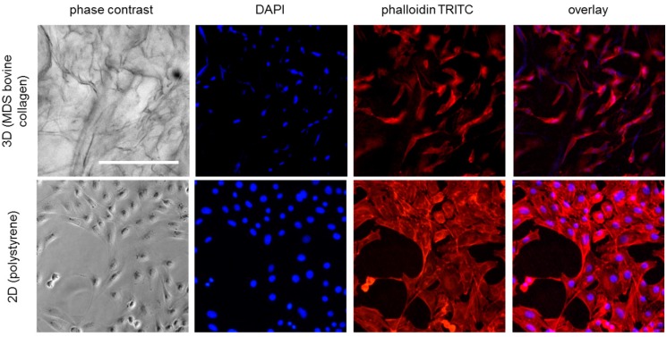
Figure 2.
Microscopic images of NIH 3T3 fibroblasts in standard 2D culture on polystyrene surface and in 3D culture on MDS bovine collagen matrix (48 h incubation; 35,000 cells/cm² seeding density). Cells were stained with DAPI (blue, nuclei) and TRITC-conjugated phalloidin (red, f-actin). Scale bar represents 250 µm.

