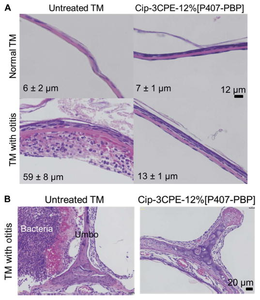
Fig. 5. In vivo effect on tissue for Cip-3CPE-12%[P407-PBP].
(A) Representative photomicrographs of hematoxylin and eosin (H&E)–stained sections of TM cross-sections in healthy TMs and TMs after 7 days of OM without or after treatment with Cip-3CPE-12%[P407-PBP]. Scale bar, 12 μm. Labeled on each panel is the TM’s average thickness ± SD (n = 4). (B) H&E-stained cross-sections of the umbo-malleus region after 7 days of OM, without or after treatment with Cip-3CPE-12%[P407-PBP]. Scale bar, 20 μm. Thickness measurements are provided in table S4.

