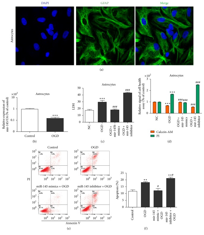
Figure 1.
miR-145 protected astrocytes from OGD-induced injury. (a) GFAP/DAPI staining of primary astrocytes (×200 magnification). (b) qRT-PCR detection of miR-145 expression in OGD primary cultured astrocytes or NC; U6 was used as the internal control (∗∗∗p < 0.001 versus control). (c) ELISA of LDH levels in astrocyte culture supernatant (###p < 0.001 versus OGD; ∗∗∗p < 0.001 versus control). (d) Calcein-AM/PI staining cell health assay. PI-positive cells were dead cells; calcein-AM-positive cells were healthy cells (###p < 0.001 versus OGD; ∗∗∗p < 0.001 versus control). (e) Flow cytometry assay of astrocyte apoptosis assay. Cells in the B2 and B4 quadrants represent apoptotic cells. (f) The percentage of apoptotic cells in each group. ∗p < 0.05 versus NC group; #p < 0.05 versus OGD group; ∗∗p < 0.01 versus control.

