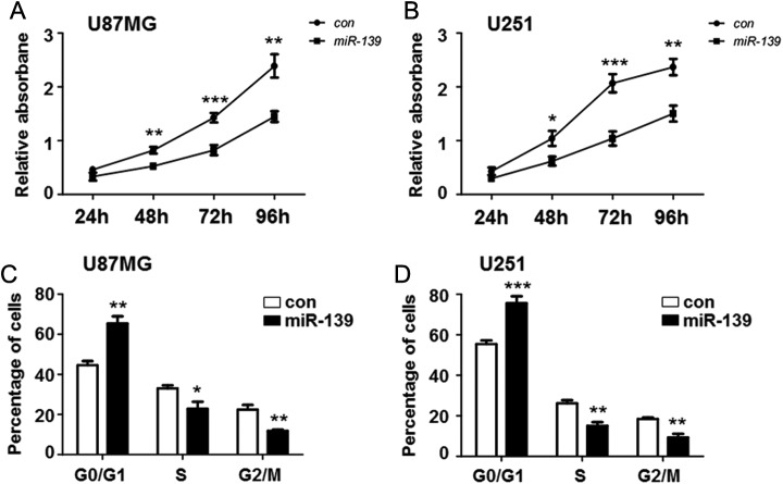
Figure 2.
miR-139 repressed gliomas proliferation. A and B, U251 and U87MG cells were seeded in 96-well plates after transfecting miR-139 or control oligonucleotide. The cell proliferation was evaluated at 24, 48, 72, and 96 hours (n = 5). C and D, U251 and U87MG cells were overexpressing miR-139 as above and collected for further flow cytometry analysis to detect the cell cycle (n = 4). Bars represent means ± SD, *P < .05, **P < .01, ***P < .001.

