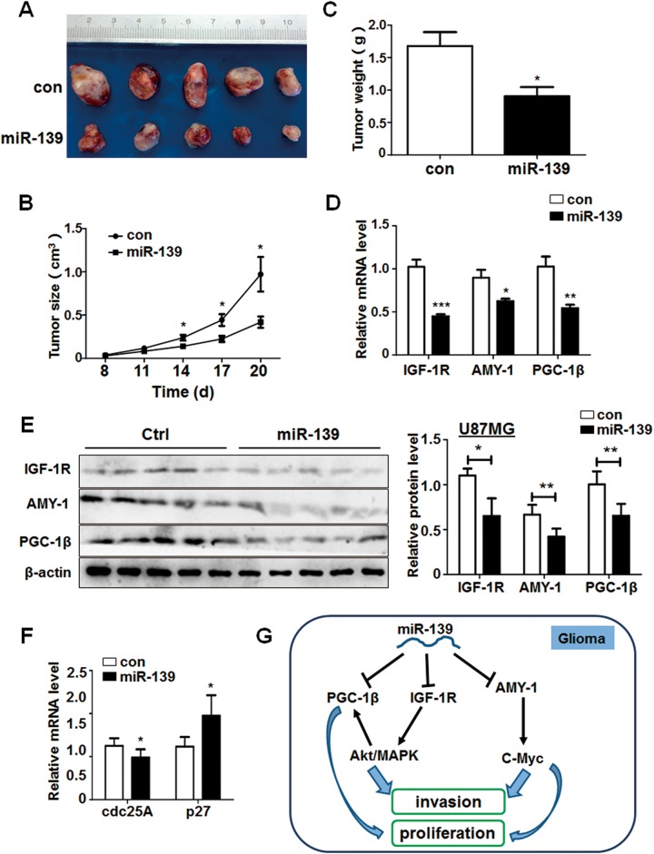
Figure 8.
miR-139 plays an antitumor function and inhibit the Akt signaling in vivo. A, U87MG cells stably expressing miR-139 and negative control were injected subcutaneously in 2 groups, respectively. The phenotype of tumors was observed after 20 days (n = 5). B and C, The growth of tumors was monitored by measuring tumor size every 3 days from the 8th day, and the tumors’ weight was measured after the mice were killed. D and E, The RNA and protein were extracted from the tumors of different groups, and the expression of IGF-1 R, associate of Myc-1 (AMY-1), and peroxisome proliferator-activated receptor γ coactivator 1β (PGC-1β) were detected. F, The RNA levels of c-Myc pathway downstream genes were determined. G, The schematic diagram of miR-139 modulated gliomas progression. Bars represent means ± SD, *P < .05, **P < .01, ***P < .001.

