
Figure 3.
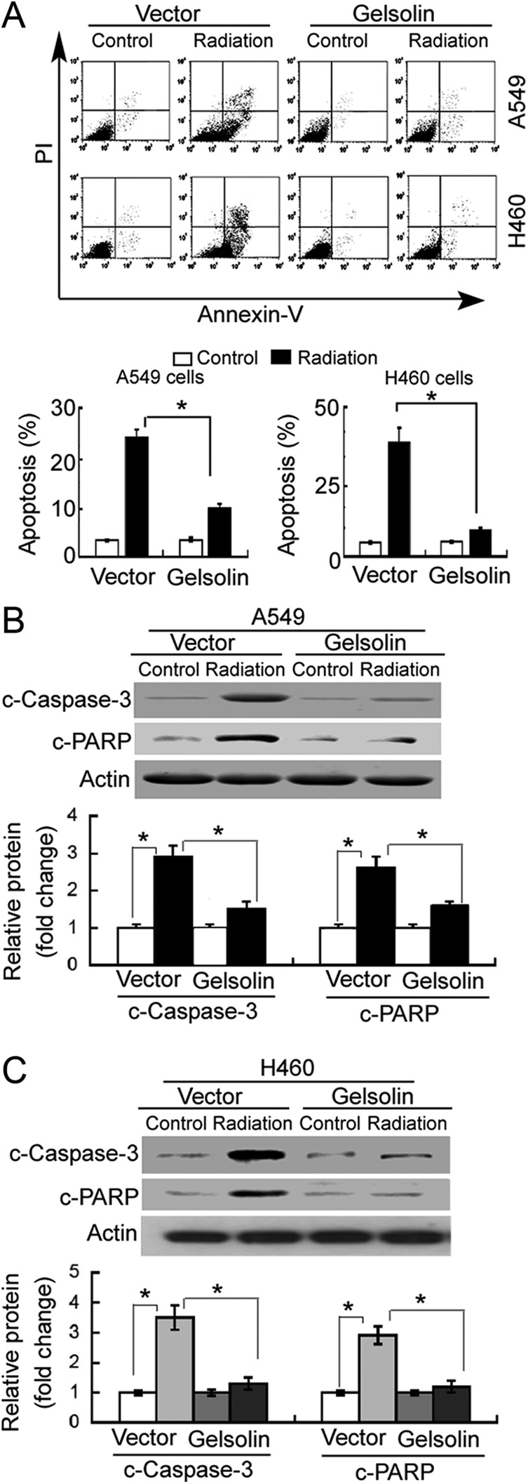
Gelsolin confers resistance to irradiation-induced apoptosis. A549 and H460 cells transfected with empty vector or gelsolin-expressing plasmid were nonirradiated (control) or exposed to 8-Gy X-ray. A, Apoptosis detected by annexin-V/propidium iodide (PI) staining and flow cytometry analysis. Representative flow cytometric dot plots showing apoptotic cells (top panels). Bar graphs (bottom panels) represent quantification of total apoptotic cells (annexin-V+/PI− or annexin-V+/PI+) from 3 independent experiments. Western blot analysis of cleaved caspase-3 (c-caspase-3) and cleaved poly adenosine diphosphate-ribose polymerase(c-PARP) proteins in (B) A549 and (C) H460 cells with indicated treatments. Bar graphs (bottom panels) represent means ± standard deviation (SD) from 3 independent experiments. *P < .05.