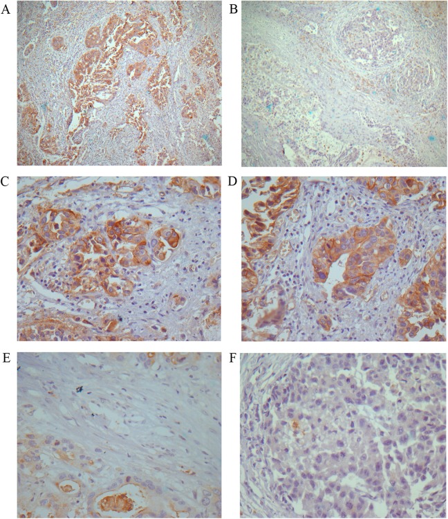
Figure 2.
Expression of aquaporin 1 (AQP1) in hilar cholangiocarcinoma (hilCC). A and B, The histological appearance of strong and low AQP1 expression, respectively (3,3′-diaminobenzidine [DAB]; magnification, ×10). C and D, The strong AQP1 expresser group (DAB; magnification, ×40). E and F, The low AQP1 expresser group (DAB; magnification, ×40).

