Figure 5.
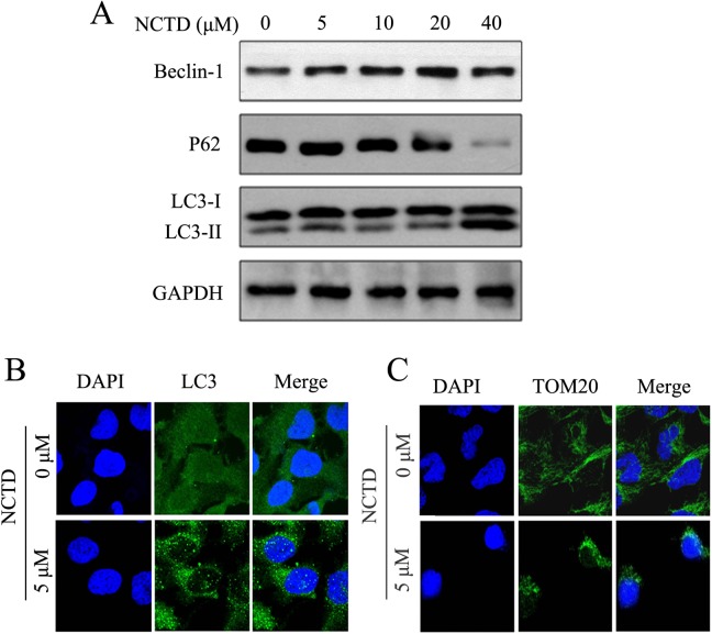
Norcantharidin (NCTD) induced autophagy and mitophagy in SK-N-SH cells. A, The expression of beclin-1, LC3, and p62 were performed by Western blotting. SK-N-SH cells were treated with NCTD (0, 5, 10, 20, or 40 μmol/L) for 24 hours. B, Punctuates of LC3 proteins in NCTD-induced SK-N-SH cells. Cells were incubated with NCTD (0 or 5 μmol/L) for 24 hours and then stained with the anti-LC3 antibody. C, Norcantharidin induced mitophagy in SKHSH cells. Different concentration of NCTD (0 or 5 μmol/L) treated SK-N-SH cells for 24 hours and stained with the anti-TOM20. Cells were examined by fluorescence confocal microscopy. Green indicates fluorescein isothiocyanate (FITC)-labeled LC3 or TOM20, and blue indicates 4′,6-diamidino-2-phenylindole dihydrochloride (DAPI)-labeled nucleus. Magnification: ×400.

