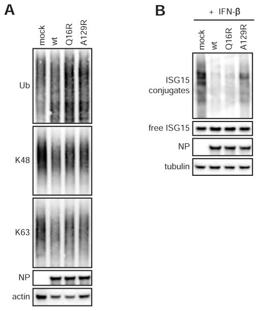
Figure 5. General levels of ubiquitinated/ISGylated proteins in CCHFV-infected cells.
(A) Western blot of ubiquitinated and ISGylated proteins in CCHFV-infected cells. Huh7 cells were infected with CCHFV at an MOI of 5. Cell lysates were harvested 24h post infection, separated by SDS-PAGE and probed for total Ub or K48- and K63-linked poly-Ub chains. (B) To assess protein ISGylation in infected cells, IFN β (1000 IU/ml) was added to the cell culture media 4 h post infection. Conjugation of ISG15 was visualized using western blotting.

