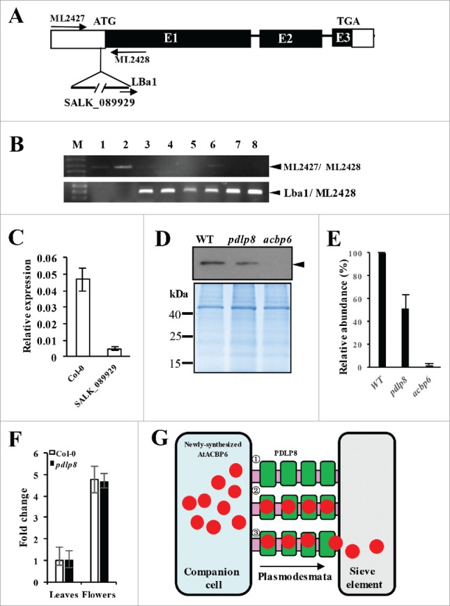
Figure 3.

Characterization of the pdlp8 mutant by PCR, qRT-PCR, and western blot analysis on its phloem exudates using anti-AtACBP6 antibodies. (A) Location of the T-DNA insertion in the PDLP8 gene. (B) Specificity of primer combinations, ML2427 and ML2428 (top gel) and LBa1 and ML2428 (bottom gel) in PCR to identify pdlp8 (homozygous) lines. Lanes 1 and 2 are from wild type 4-week-old rosette leaf DNA. Lanes 3 to 8 are from pdlp8 mutant plant DNA. The 0.65-kb ML2427/ML2428, and 0.3-kb Lba1/ML2428 PCR bands are denoted by arrowheads. (C) qRT-PCR analysis of PDLP8 expression in Col-0 and the SALK_089929 (pdlp8 mutant). Relative expression level of PDLP8 was normalized against ACTIN2. (D) Western blot analysis on 5-week-old phloem exudates of Col-0, pdlp8 and acbp6 using anti-AtACBP6 antibodies. The 10.4-kDa AtACBP6 band is indicated by an arrowhead. Coomassie Blue-stained gel identically loaded with proteins as in western blot analysis displayed below. (E) The immunoreactive bands of the total phloem extract proteins were densitometrically quantified. Each value represents the mean of three replicates (± SE). (F) qRT-PCR analysis on AtACBP6 mRNA expression in 5-week-old leaves and flowers from Arabidopsis Col-0 and pdlp8. The AtACBP6 mRNA expression in 5-week-old leaves from Arabidopsis Col-0 was normalized as 1, and fold changes in AtACBP6 mRNA expression in pdlp8 were compared. (G) Previous studies have demonstrated the AtACBP6 protein could be transported from companion cells to sieve elements via the plasmodesmata.28 A model indicating the role for AtACBP6-PDLP8 interaction in influencing AtACBP6 transport from the companion cell (blue) to the sieve element (grey) is proposed.1 PDLP8 (green) is localized to the plasmodesmata (pink).2 AtACBP6 (red) presumably interacts with PDLP8.3 PDLP8 (green) may facilitate AtACBP6 transport from the companion cells (blue) to the sieve element (grey), by unloading AtACBP6 (red) to the sieve element.
