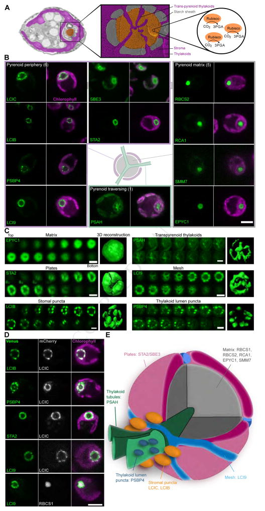
Figure 4. Pyrenoid Proteins Show at Least Six Distinct Localization Patterns and Reveal Three New Protein Layers.
(A) A false-color transmission electron micrograph and deep-etched freeze-fractured image of the pyrenoid highlight the pyrenoid tubules, starch sheath and pyrenoid matrix where the principal carbon fixing enzyme, Rubisco, is located. Images courtesy of Moritz Meyer, Ursula Goodenough and Robyn Roth.
(B) Proteins showing various localization patterns within the pyrenoid are illustrated. Scale bar: 5 μm.
(C) Confocal sections distinguish different localization patterns within the pyrenoid. Each end panel is a space-filling reconstruction. Scale bars: 2 μm.
(D) Dual tagging refined the spatial distribution of proteins in the pyrenoid. Scale bar: 5 μm.
(E) A proposed pyrenoid model highlighting the distinct spatial protein-containing regions.

