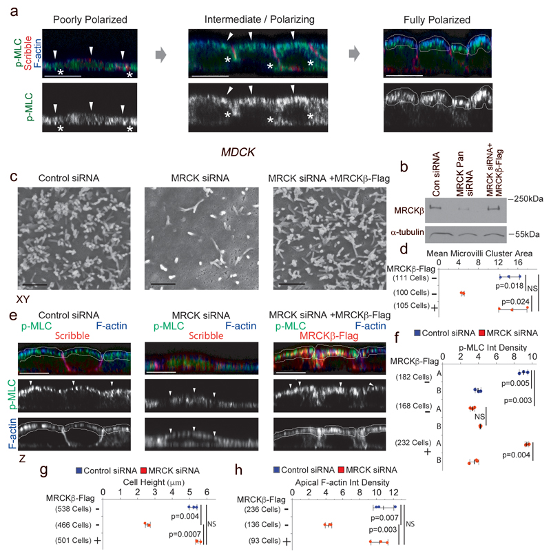
Figure 1. MRCK activates apical actomyosin contractility that controls apical morphogenesis.
(a) Spontaneous polarization of MDCK cells leads to the formation of apical actomyosin caps positive for p-MLC and F-actin. (b) Expression of MRCKβ as analysed by immunoblotting of total cell extracts. (c,d) Scanning electron microscopy of apical domains reveals levels of microvilli induction by MDCK cells upon depletion of MRCK without or with complementation with exogenously expressed MRCKβ-flag. (e,f) Measurement of active Myosin at cortical caps (A) and basal membrane (B) during polarization and differentiation of cells depleted of or rescued for MRCK expression by confocal immunofluorescence microscopy. (g,h) Measured cell height and F-actin levels in polarizing cells with or without MRCK function. For all quantifications, n=3 independent experiments and shown are the data points, means ± 1 SD (in black), the total number of cells analysed for each type of sample across all experiments, and p-values derived from t-tests. Arrowheads point to the apical cortex demarked by F-actin. Unprocessed original scans of blots are shown in Supplementary Figure 8. Scale bars: electron micrographs, 1μm; confocal immunofluorescence images, 10μm.

