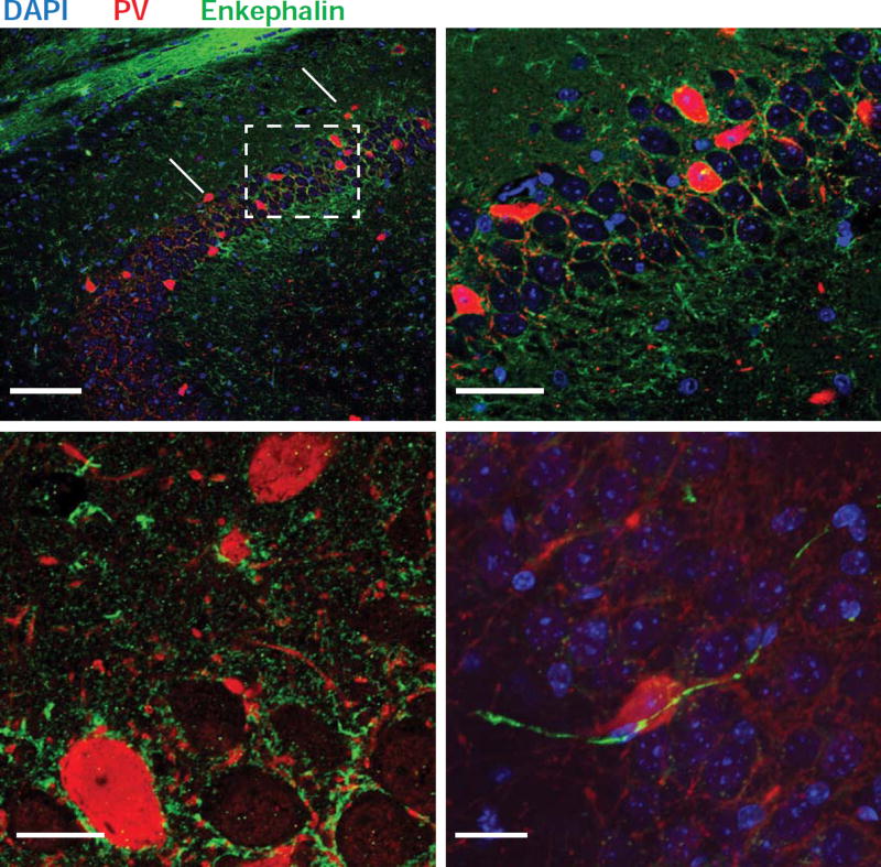
Figure 7. ENK+ fibers contact PV+ INs.
A–C. Immunohistochemistry of the hippocampus with antibodies recognizing PV (red) and enkephalin (green). Fitzgerald enkephalin antibody used in A and B; Santa Cruz enkephalin antibody used in C; see Experimental Procedures. Arrowhead in A1 points to the end of the mossy fiber pathway. A2. Higher magnification view of dashed box region in A1. B. High magnification view of the tight network of ENK+ (green) and PV+ fibers (red) surrounding unstained CA2 PNs somas (asterisks). Two PV+ neuron somas are also seen (large red somas). In C, an ENK+ fiber can be seen impinging on a PV+ soma.

