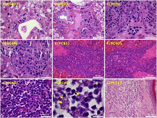
Fig 3. Histopathological changes in affected tissues of control (images A, C, E, I) and vaccinated (images B, D, F-H) dogs naturally infected with Leishmania infantum.
Photomicrographs sections from the liver of dogs with subclinical infections showing minimal mononuclear cell infiltration in portal spaces (A-B), as well as intralobular granulomas containing activated but parasite-free macrophages, surrounded by plasma cells and occasional lymphocytes (C-D). Also illustrated are photomicrographs of sections from spleen (E-G), lymph node ((H) and ear skin (I) showing a more marked mononuclear cell infiltration, particularly composed of plasma cells, activated macrophages, including parasite-containing phagocytes (arrows), thus confirming the sustained course of infection. The number within parenthesis indicates dog code. Images were obtained from paraffin-embedded sections stained with haematoxylin-eosin at different magnification.

