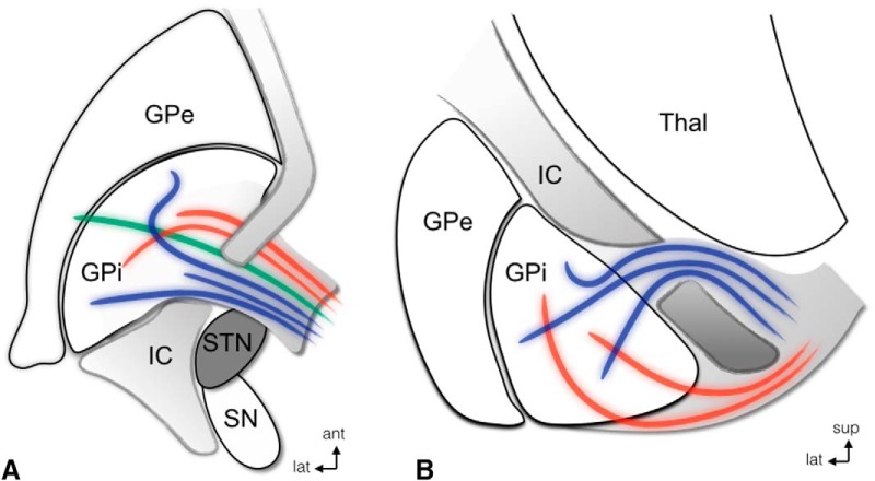
Figure 3.
STN and pallidofugal fibers. Axial (A) and coronal (B) schematic representations of the AL (red) and LF (H2; blue), in relationship to the STN, in nonhuman primates. Note that both the tracts travel dorsal to the most anterior aspect of the STN. A, The thalamic fasciculus is represented in green. ant, anterior; lat, lateral; sup, superior. Modified and reprinted with permission from Parent and Parent (2004).

