Fig. 1.
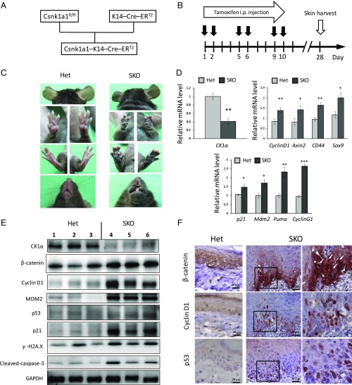
CK1α ablation in keratinocytes induces skin hyperpigmentation and activation of Wnt and p53 signaling pathways. (A) Scheme of SKO mice crossing. (B) CK1α is ablated in keratinocytes by i.p. tamoxifen injection. All samples were harvested at day 28. (C) SKO mice show skin hyperpigmentation of ears, paws, mouth, and trunk at 1 mo compared with heterozygous control (Het) mice. (D) RT-PCR analyses of the epidermis. Expression of CK1α mRNA is down-regulated in SKO mice. The mRNAs of Wnt-target genes and p53-target genes are up-regulated in SKO mice. *P < 0.05, **P < 0.01, and ***P < 0.001. (E) Western blot analyses of the epidermis. The reduced level of CK1α protein causes β-catenin stabilization and increases levels of cyclin D1, p53, p21, and Mdm2 in SKO mice. Increased levels of markers for DNA damage response (γ-H2AX) and apoptosis (cleaved-caspase 3) are also detected in SKO mice. (F) Immunohistochemistry of paw skin. Positive nuclear staining of β-catenin, Cyclin D1, and p53 in the basal layer of the epidermis of SKO mice. Panels in the Right column are 2.48× magnified on the square in the Middle column. (Scale bars, 20 μm.)

