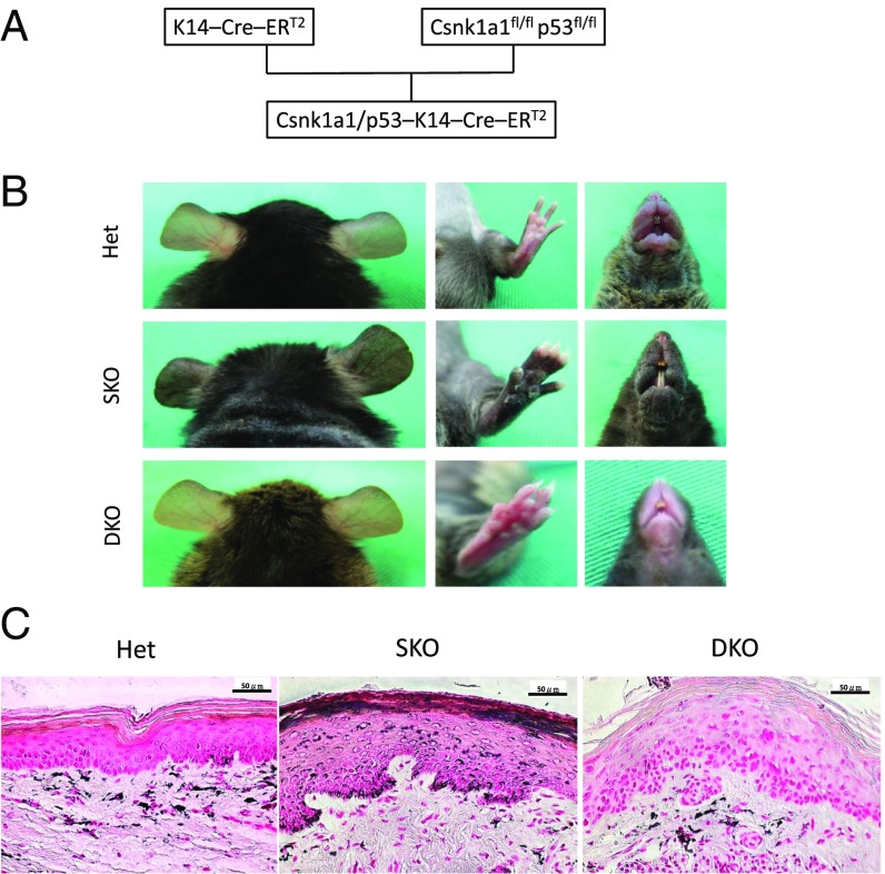
Fig. 3.
CK1α ablation in keratinocyte-induced skin hyperpigmentation is p53-dependent. (A) Scheme of the DKO mice crossing. (B) SKO mice develop skin hyperpigmentation, but DKO mice do not show skin hyperpigmentation. (C) Fontana–Masson staining of the paw skin. Melanin distribution in heterozygous control (Het) mice is mainly located in the dermis and less in the epidermis. In SKO mice, the epidermis becomes hyperplastic with increased melanin deposition. In DKO mice, the epidermis becomes dysplastic without melanin deposition. (Scale bars, 50 μm.)

