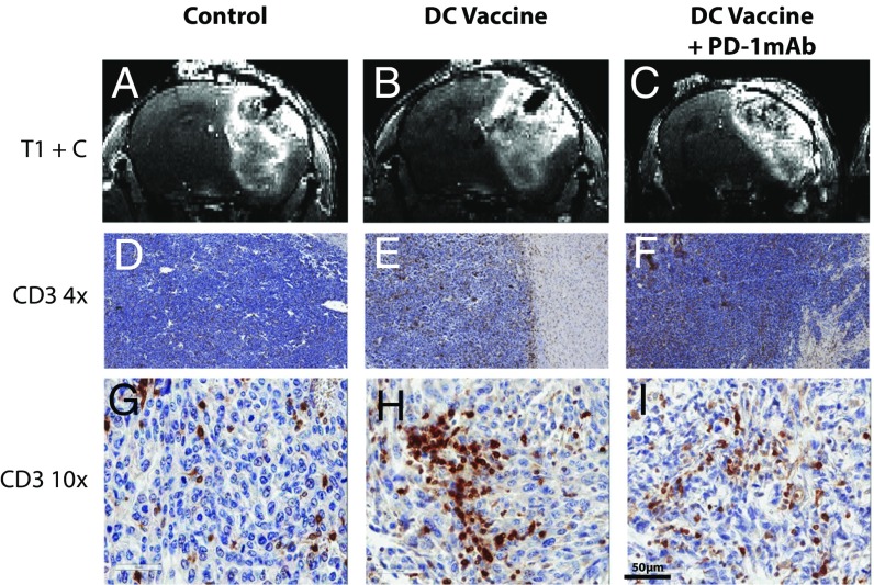
Fig. 1.
Standard contrast-enhanced MRI cannot distinguish tumor growth from pseudoprogression in glioma-bearing mice. (A–C) Representative coronal sections of T1-weighted MRI images following i.v. injection of contrast agent. (D–I) 4× magnified (D–F) and 10×- magnified (G–I) cross-sections of mice from control, DCVax, and DCVax + PD-1 mAb mice after immunohistochemical staining for CD3. Images were obtained from representative mice in experiments repeated multiple times; n = 4–6 mice per group.

