Fig. 1.
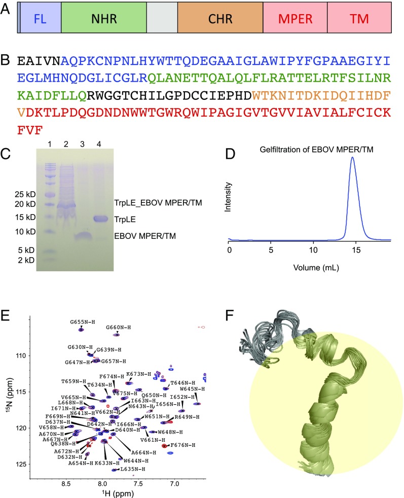
Expression, purification, and solution NMR structure of the EBOV MPER/TM domain. (A) EBOV GP2 domain structure. CHR, C-heptad repeat region (orange); FL, fusion loop (blue); MPER, membrane proximal external region (red); NHR, N-heptad repeat region (green); TM, transmembrane region (red). (B) Primary structure of EBOV GP2 (Zaire strain). Color codes are matched with domain structure. (C) SDS/PAGE gel showing the isolation of the MPER/TM domain from the expression fusion protein construct with the Trp leader protein. Lane 1, markers; lane 2, expressed fusion protein after solubilization from inclusion bodies; lane 3, cleaved MPER/TM domain; lane 4, Trp leader protein. (D) Size exclusion chromatography of the MPER/TM domain in DPC micelles (20 mM phosphate, 100 mM NaCl, 0.2% DPC) at pH 7.0 showing a single homogeneous symmetric peak. (E) HSQC spectra of the MPER/TM domain in DPC micelles at pH 7.0 (blue) and pH 5.5 (red). All backbone atoms were assigned as shown. (F) Twenty lowest energy structures of the MPER/TM domain in DPC micelles at pH 5.5.

