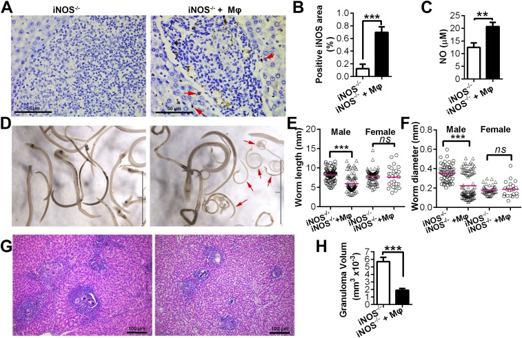
Fig. S6.
Adoptive transfer of WT macrophages into infected iNOS−/− rats. Adoptive transfer of WT macrophages was performed in iNOS−/− rats, as described in SI Materials and Methods (group iNOS−/− + Mφ), simultaneously with a group of infected iNOS−/− rats that did not receive macrophages; instead, PBS was used as an additional control (group iNOS−/−). The rats were killed on day 43 (6 wk postinfection). (A) Expression of iNOS in liver was identified by immunohistochemistry using an iNOS antibody. Arrows indicate the iNOS signal. (B) Quantitation of the positive area of fields of view showing iNOS expression. (C) NO concentration in the serum of infected iNOS−/− rats and iNOS−/− recipients at 6 wk postinfection. (D) Representative micrographs showing parasites present. Arrows identify stunted parasites. (E and F) Worm lengths and diameters were measured from digital micrographs. Mean values are represented by horizontal bars. (G) H & E stain of representative hepatic granulomas. (H) Quantitation of hepatic granuloma sizes as measured from H&E-stained slides. Data are expressed as the mean ± SEM from two biological repeats (n = 10). **P < 0.01; ***P < 0.001. ns, not significant. Scale bars represent 50 μm (A), 5 mm (D), or 100 μm (G), respectively.

