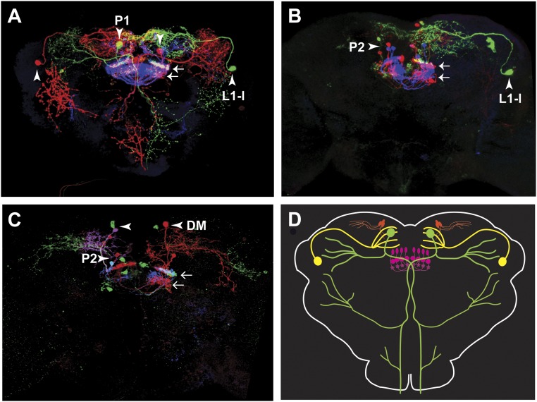
Fig. S8.
Neuroanatomy of neurons expressing NPF-GAL4. Representative multicolor flip-out (MCFO) results, showing the different types of NPF-GAL4 neurons: large dorsal medial (P1) neuron (A); large dorsal lateral (L1-l) neuron (A and B); small fan-shaped body (P2) interneurons (B and C); small dorsal medial (DM) neuron. (D) Schematic representation of the different subtypes of neurons expressing NPF-GAL4. In all images, small arrows indicate the projections from P2 neurons to the fan-shaped body, while big arrowheads indicate the neuron of interest in each panel. The male-specific neurons L1-s, D1, and D2 (20) were rarely labeled and have therefore been omitted. (Magnification: 20×.)

