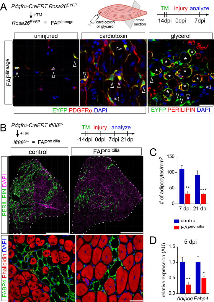
Figure 2. Removing FAP cilia impairs fat formation.
(A) Detection of lineage label (EYFP, green) in FAPs (arrowheads; PDGFRα, red) 7 dpi with cardiotoxin injection and in adipocytes (asterisks; PERILIPIN, red) 7 dpi with glycerol injection using PDGFRα-CreERT Rosa26EYFP mice. Scale bar is 25 µm.
(B) Immunofluorescence for adipocytes (PERILIPIN, top, green or FABP4, bottom, red) 21 dpi with glycerol injection after conditional removal of FAP cilia (n=8–12 mice per genotype). Nuclei are stained with DAPI. Myofibers are stained with phalloidin (red). Scale bars are 1 mm (top) and 25 µm (bottom).
(C) Quantifications of the number of adipocytes present per 1 mm2 of injured area 7 dpi (n=4 per genotype) or 21 dpi with glycerol injection (n=8–12 per genotype).
(D) RT-qPCR for mature adipocyte markers (Adipoq and Fabp4) from whole muscle RNA 5 dpi with glycerol injection (n=4 per genotype). All data are represented as mean ± SEM. See also Figure S2.

