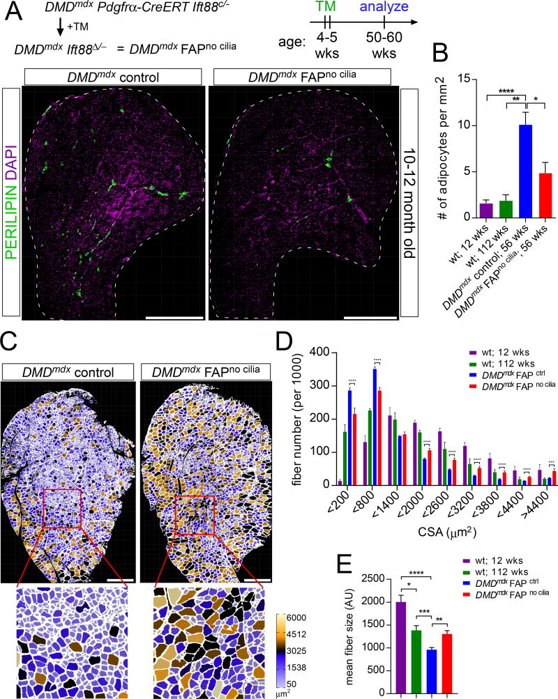
Figure 3. Loss of FAP cilia reduces fatty degeneration of Dmdmdx muscle.
(A) Immunofluorescence for adipocytes (PERILIPIN, green) after conditional removal of FAP cilia in 10–12 month old DMDmdx mice. Scale bar is 1 mm.
(B) Quantifications of the number of adipocyte present per 1 mm2 of tibialis anterior muscle in 12 week old wild type mice (n=10), 2 year old wild type mice (n=5), 1 year old DMDmdx control mice (n=17) and DMDmdx FAPno cilia mice (n=8).
(C) Myofibers of 10–12 month old DMDmdx FAPno cilia and DMDmdx control mice color-coded based on size of cross-sectional area using the ROI Color Coder function in ImageJ (see Experimental Procedures for details). Scale bar is 0.5 mm.
(D) Distribution of the number of fibers based on their cross-sectional area in 12 week old (n=7) and 2 year old (n=5) wild type and DMDmdx mice with cilia (n=11) and no cilia (n=5).
(E) Average cross-sectional area size of myofibers from wild type (n=7 for 12 week old and n=5 for 2 year old), DMDmdx FAPno cilia (n=5) and DMDmdx control mice (n=11). All data are represented as mean ± SEM. See also Figure S3.

