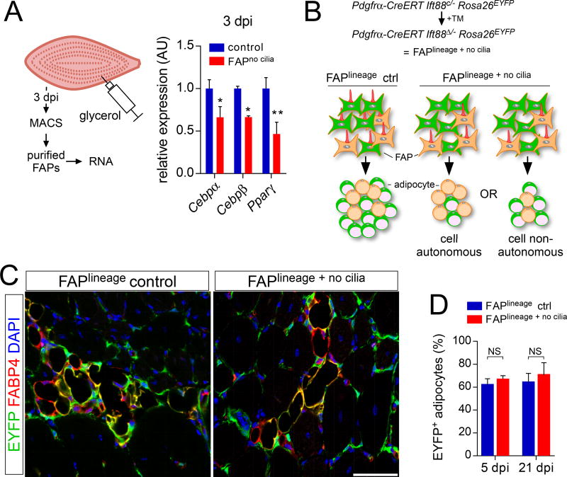
Figure 5. Ciliary Hh signaling reduces adipocyte differentiation via a cell non-autonomous mechanism.
(A) RT-qPCR analysis of MACS-purified FAPs of control and FAPno cilia mice 3 dpi with glycerol injection (n=5–6 per genotype) for the early adipocyte genes Cebpα, Cebpβ and Pparγ.
(B) Combining the Rosa26EYFP allele with Pdgfrα-CreERT Ift88c/− mice allows for simultaneous deletion of cilia and activation of the lineage mark. A specific reduction in the percentage of EYFP+ adipocytes (green) would argue for a cell autonomous effect after loss of FAP cilia, whereas a reduction in total number of both EYFP+ and EYFP− adipocytes (without affecting the percentage expressing EYFP) would suggest a cell non-autonomous mechanism.
(C) Immunofluorescence for lineage marker (EYFP, green) and adipocytes (FABP4, red) after conditional removal of IFT88 in FAPs 21 dpi with glycerol injection. Scale bar is 50 µm.
(D) Quantifications of the percentage of EYFP-expressing FAPs at 5 dpi (n=4–5 per genotype) and 21 dpi (n=5–6 per genotype) with glycerol injection. All data are represented as mean ± SEM. See also Figure S5.

