Figure 1.
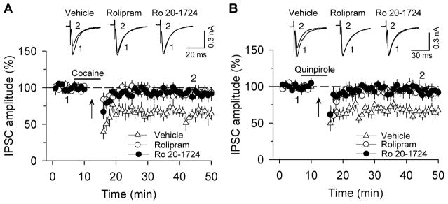
Selective PDE4 inhibitors rolipram and Ro 20-1724 blocked I-LTD in VTA dopamine neurons. (A) The presence of cocaine (3 μM; indicated by horizontal bar) during the 10 Hz stimulation (indicated by arrow “↑”) induced I-LTD in VTA dopamine neurons (n = 6). This I-LTD was blocked by PDE4 inhibitors rolipram (1 μM; n = 8; p < 0.05 vs. control) and Ro 20-1724 (200 μM; n = 8; p < 0.05 vs. control). The PDE4 inhibitors were present throughout the whole-cell recordings. Sample IPSCs before (indicated by “1”) and after (indicated by “2”) the 10 Hz stimulation are shown on the top. (B) The presence of D2 receptor agonist quinpirole (1 μM) during the 10 Hz stimulation induced I-LTD in VTA dopamine neurons (n = 7). This I-LTD was blocked by rolipram (1 μM; n = 8; p < 0.05 vs. control) or Ro 20-1724 (200 μM; n = 7; p < 0.05 vs. control). Error bars indicate SEM (used with the permission of Neuropsychopharmacology).

