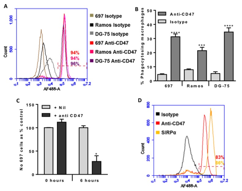
Figure 1. CD47 is expressed on the surface of malignant B-lymphocytes, and anti-CD47 antibodies increase their phagocytosis by macrophages dramatically reducing their numbers within 6 hours.
A. Antibody binding to 697 (pre-B lymphoblast cell line), Ramos and DG-75 (Burkitt lymphoma mature B lymphocyte cell lines) incubated with 10 µg/mL of isotype control or B6H12 anti-CD47 antibody, then AF488-conjugated secondary before analysing by flow cytometry. Representative example of 3 experiments. The % of each cell type binding the anti-CD47 antibody is given, and was gated on live cells (propidium iodide negative). B. U937 cells were matured into macrophages with 10nM PMA for 24 hours and further 48 hours incubation. Target B cells were stained with TAMRA, and macrophages with CFSE before co-incubating for 2 hours at 1:1 ratio with isotype or anti-CD47 B6H12 at 2 µg/mL. Phagocytosis assessed by measuring % of double positive macrophages. N≥4. Bars are mean ± SEM, *** / **** p < 0.001 / 0.0001, compared to isotype for that cell line. C. Phagocytosis assay prepared as above, but 697 stained with JC-1 and U937 with Höechst 33342. The number of free, live 697 cells was counted at 0 and 6 hours after addition of anti-CD47 antibody or nil. N = 4. D. Antibody binding to U937-derived macrophages incubated with 10 µg/mL of isotype control, B6H12 anti-CD47 antibody or anti-SIRPα antibody, then AF488-conjugated secondary before analysing by flow cytometry. Representative example of 3 experiments. The % of cells binding the anti-CD47 antibody and anti-SIRPα antibody are given, and was gated on live cells (propidium iodide negative). N = 3. Bars are mean ± SEM, * p < 0.05 compared with control.

