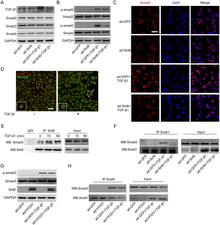
Figure 4. Sirt6 attenuates TGF-β1/Smad3 signaling.
(A) A549 cells were transfected with ad-GFP or ad-Sirt6 in the absence or presence of TGF-β1 (5 ng/ml) for 24 h, and the protein levels of TGF-β1, Smad2, Smad3, and Smad4 were measured by Western blot. (B-C) A549 cells were transfected with ad-GFP or ad-Sirt6 in the absence or presence of TGF-β1 (5 ng/ml) for 60 min. (B) The protein levels of p-Smad2 and p-Smad3 were measured by Western blot. (C) Representative images of the immunofluorescent staining of Smad3. Scale bar, 40 μm. (D) Representative images of double immunostaining of Sirt6 and Smad3. Scale bar, 50 μm. (E) A549 cells were treated with or without TGF-β1 (5 ng/ml) for 15 and 60 min. Total protein was subjected to co-IP with anti-Sirt6 antibody. (F) A549 cells were transfected with ad-GFP or ad-Sirt6 in the absence or presence of TGF-β1 (5 ng/ml) for 24 h, followed by co-IP with anti-Snail1 antibody. A549 cells were transfected with ad-GFP or ad-H133Y in the absence or presence of TGF-β1 (5 ng/ml) followed by (G) Western blot to measure the protein levels of p-Smad3 after 60 min treatment and (H) co-IP with anti-Snail1 antibody after 24 h treatment.

