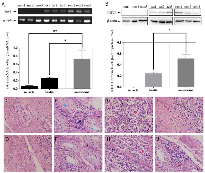
Figure 1. KIFC1 is enriched in human seminoma samples in both mRNA and protein level.
(A) Comparison of mRNA expression level among human muscle (mus), testis (tes) and seminoma (sem) tissue samples. kifc1 mRNA is enriched in seminoma samples. (B) Comparison of protein expression level among human muscle (mus), testis (tes) and seminoma (sem) tissue samples. KIFC1 protein is enriched in both testis samples and seminoma samples, and KIFC1 protein expression level of seminoma samples is significantly higher than that of testis samples. (C-J) HE staining results of human muscle tissues (C, D) and testis tissues (E, F) near the seminoma tissue and the seminoma tissue (G, H) from seminoma patient. (I) The transition of the seminoma to the muscle tissue. (J) The invasion of the cancer cells into the testis tissue. Black arrows show infiltrating lymphocytes. Blue arrows show seminiferous tubules of normal testis tissue. Red arrows show cancer cells. Yellow arrow shows muscle tissue. In the both testis and seminoma, a number of lymphocyte infiltration with bleeding are showed. In seminoma tissues, compared with normal tissues, seminiferous tubules were disrupted and replaced by seminoma cells with loose nuclei and watery cytoplasm.

