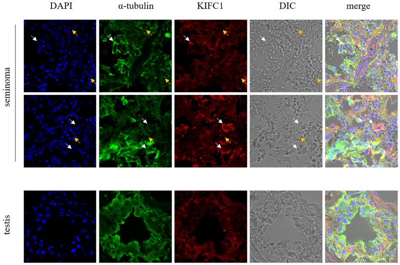
Figure 2. Localization of KIFC1 in seminoma tissue samples and nearby testis tissue samples from human determined by immuno-florescent staining.
KIFC1 is omnipresent in both seminoma samples and testis samples and co-localizes with MT. White arrows show seminoma cells with loose nuclei and watery cytoplasm, in which KIFC1 signal is strong. Yellow arrows show other cells such as lymphocytes, in which KIFC1 signal is weak. Compared with normal testis tissue, in seminoma tissues the characteristic structure of seminiferous tubules is completely disrupted, and KIFC1 has a tendency to be concentrated around the loose nuclei.

