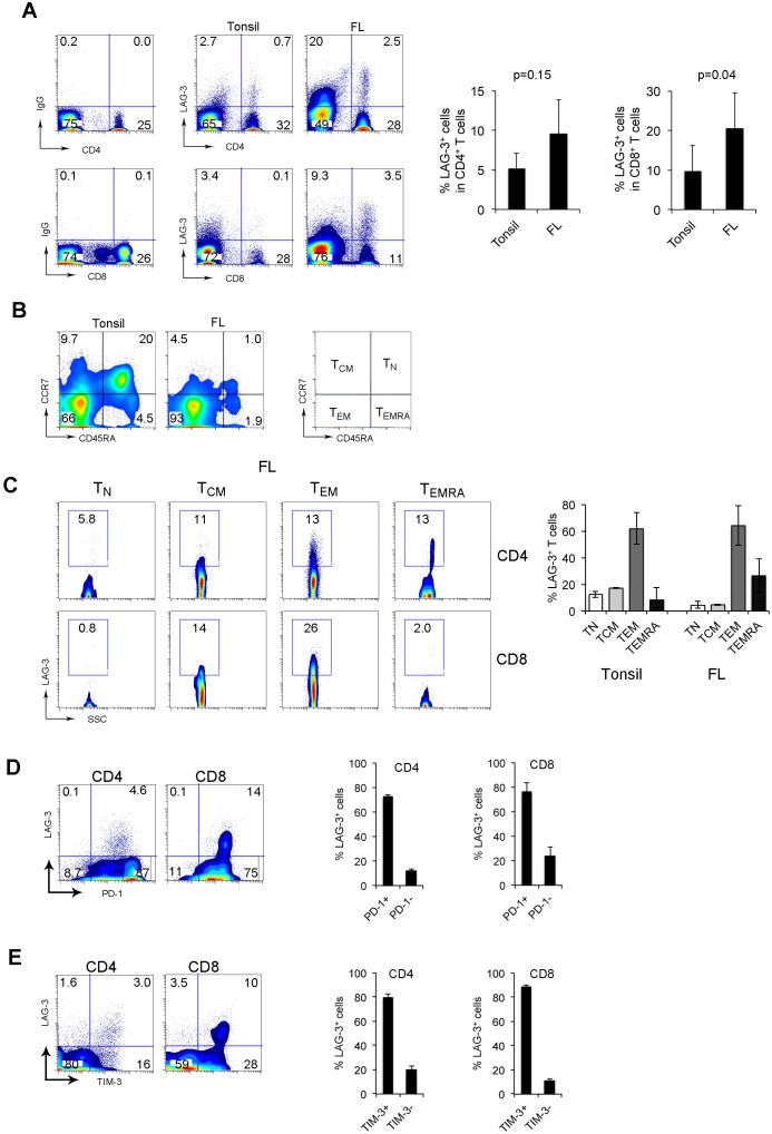
Figure 2. LAG-3 is primarily expressed on intratumoral PD-1+ T cells in FL.
(A) LAG-3 expression on CD4+ or CD8+ T cells from tonsils and FL specimens. The right graphs summarize percentages of LAG-3+CD4+ or CD8+ T cells from tonsils and FL. (B) CD45RA and CCR7 expression on CD3+ T cells from tonsils and FL specimens. TN: naïve T cells (CD45RA+CCR7+), TCM: central memory (CD45RA-CCR7+), TEM: effector memory (CD45RA-CCR7-) and TEMRA: terminally-differentiated T cells (CD45RA+CCR7-). (C) LAG-3 expression by CD4+ or CD8+ T cell subsets. The right graph summarizes percentages of LAG-3+ T cells in T cell subsets of TN, TCM, TEM and TEMRA. (n=5 for tonsils, n=3 for FL patients). (D-E) Co-expression of LAG-3 and PD-1 (D) or TIM-3 (E) in CD4+ or CD8+ T cells. The right graphs show percentage of LAG-3+ T cells in subsets of PD-1 or TIM-3 T cells (n=6).

