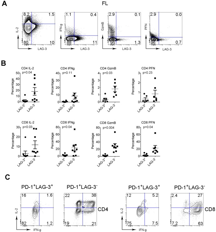
Figure 4. The function of intratumoral LAG-3+ T cells is reduced.
(A) Co-staining of IL-2, IFN-γ, GzmB and PFN with LAG-3 in intratumoral T cells from FL patients. (B) Graphs summarize percentages of cytokine- (IL-2 and IFN-γ) and granule- (GzmB and PFN) producing CD4+ or CD8+ T cells by LAG-3- or LAG-3+ subset from FL patients. (C) Expression of IL-2 and IFN-γ by PD-1+LAG-3- or PD-1+LAG-3+ T cells in CD4 or CD8 subset. PD-1+LAG-3- or PD-1+LAG-3+ T cells were defined by gating co-staining of PD-1 and LAG-3.

