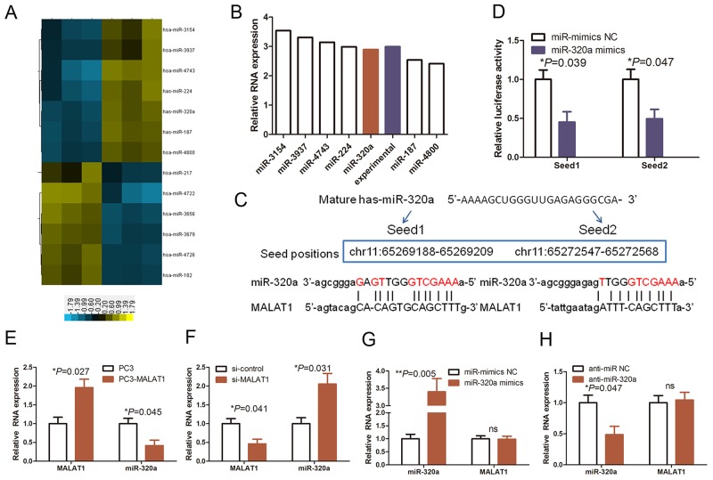
Figure 3. The reciprocal interaction of MALAT1 and miR-320a in HUVECs.
(A) The data on miRNA profiling in MALAT1 knocked down HUVECs. Our previous study assessed differentially expressed miRNAs between MALAT1 siRNA and control siRNA-transfected HUVECs using miRNA array. The shaded yellow section represents increased miRNA expression, whereas the shaded blue section represents decreased miRNA expression. (B) The graph shows the top seven upregulated miRNAs and were confirmed by qRT-PCR. (C) Prediction and alignment of the potential miR-320a binding regions to MALAT1. (D) Luciferase reporter assay. Relative luciferase activity mediated by the reporter constructs harboring the miR-320 binding site upon transfection with miR-320a mimics or NC. (E) and (F) qRT-PCR. HUVECs were grown and transfected with MALAT1 cDNA (PC3-MALAT1) or vector-only plasmid or MALAT1 siRNA (si-MALAT1) or negative control siRNA and then subjected to qRT-PCR analysis of MALAT1 and miR-320a levels. (G) and (H) qRT-PCR. HUVECs were grown and transfected with miR-320 mimics, anti- miR-320 or their NC controls and then subjected to qRT-PCR analysis of MALAT1 and miR-320a levels.

