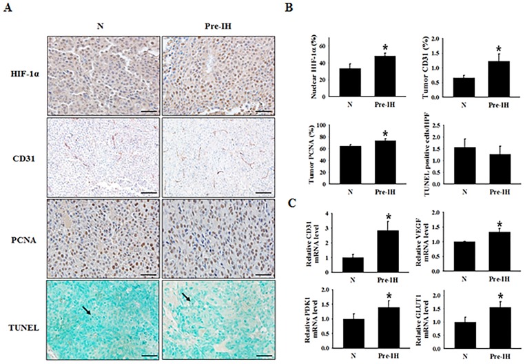
Figure 3. Expression of a cell-proliferation marker, endothelial cell markers, and hypoxia-response transcription factor in tumor tissues from mice in the no-conditioning (N) and pre-intermittent hypoxia conditioning (Pre-IH) groups.
(A) Representative immunohistochemical staining images for HIF-1α, PCNA, CD31, and TUNEL-positive nuclei. Tumor tissues were collected on day 19 post-tumor injection. (B) Quantification of the % of cells staining positive for nuclear HIF-1α, the % CD31-stained area, the % PCNA-positive cells, and the average number of TUNEL-positive cells per high-power field. (C) Relative CD31, VEGFA, PDK1, and GLUT1 mRNA-expression levels in tumors from mice in the N and Pre-IH groups. The arrow indicates TUNEL-positive nuclei. Scale bars, 50 μm except for CD31 (100 μm). *P < 0.05 compared to the N group, as determined by the Mann–Whitney U test.

