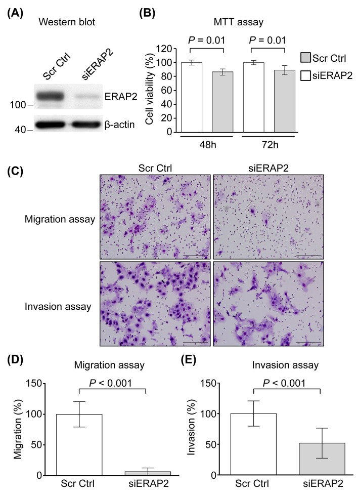
Figure 3. Involvement of ERAP2 in viability, migration, and invasion of OSCC cells.
(A) SCC4 cells were transfected with control siRNA (Scr Ctrl) and ERAP2 siRNA (siERAP2), respectively. After 48 h, protein extracts (20 μg) were prepared and ERAP2 was detected with Western blot. (B) Quantitative analysis of the MTT assay. Data are presented as mean values obtained from three independent experiments. Error bars indicated the standard deviation. (C) Representative microphotographs of filters obtained from the 16 h transwell migration (D) and 24 h invasion (E) assays. Original magnification: 200×. The migration (D) and invasion (E) assays were performed using SCC4 cells in which ERAP2 was eliminated.

